Root canal treatment — Endodontology
Maintaining teeth for a lifetime is not always easy — especially if they are inflamed. However, in the vast majority of cases, your own teeth are better than any good replacement. Root canal treatment is highly likely to achieve this.
On this page we would like to explain the most important aspects of root canal treatment.
Our specialists for endodontology

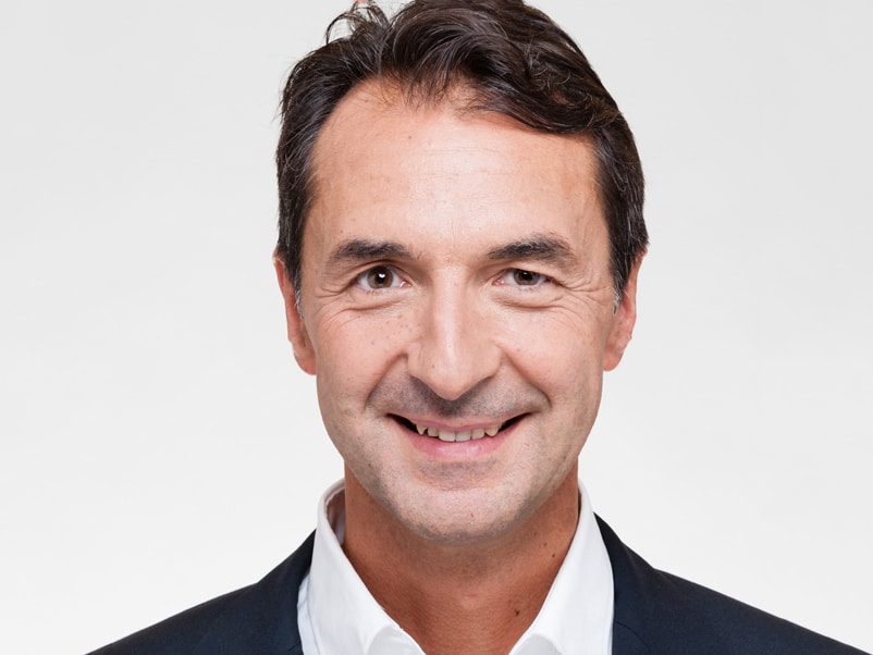
Dr. Marco Georgi, M.Sc., is a specialist in microscopic endodontology, endodontic retreatments and microsurgery, and a popular lecturer in many training institutions. For more than 10 years he has also taught internationally and has been appointed as Scientific Director of Endodontic Education in the State Chamber of Physicians of Hesse. He regularly leads training events at the PRAXIS AM KURECK for other dentists and endodontologists. He is the founding President of the Association of Certified German Endodontologists


Why is my tooth bad?

In most cases a tooth decays due to caries-causing bacteria but accidents and dental or orthodontic treatments can also be a cause. Inflammation or infection within the tooth can result from these stimuli. Inside a tooth there is a branching system of canals containing living tissue (pulp) with nerves and blood vessels. Unfortunately the defensive abilities of this tissue are very limited so the body sometimes cannot sufficiently deal with the irritation and heal the infection.
There used to be no way of saving these teeth. Because the canal system in a tooth is often very fragile and has lots of bends, there was no possibility of treatment so the tooth had to be removed.
What can the dentist do so that I can keep my tooth?
In order to be able to preserve an inflamed or dead tooth its root must be treated. The bacteria in the tooth must be removed and the tooth sealed so that no new germs can get in.
Because the root system of a tooth has lots of small branches, like a tree (some as small as 0.06mm!), they can only be seen under a microscope. This technology and the associated flexible, minute instruments enable the optimum preparation for removal of the bacteria and the diseased tissue – and therefore also increased treatment success. Then the residue is removed with a rinsing solution.
As a final preparation for filling the root canal system, the root canal is prepared with highly flexible micro-files. For the filling, the material gutta-percha, which is related to natural rubber, is heated and put into the now perfectly prepared system in combination with an adhesive cement.
Only if the root system can be thoroughly cleaned of the bacteria, the infection can be eliminated and the bone can heal again.
What are the chances of preserving my tooth?
Obviously this depends on the individual circumstances, such as the degree of inflammation and your mouth hygiene. But the anatomy of the tooth and perhaps other factors such as the loss of periodontal apparatus through the formation of cysts play a considerable role.
The good news: The results of long-term studies in the USA and Scandinavia have shown that professional endodontic treatment using the above methods has a success rate of over 80%
How much does the treatment cost?
The cost of the treatment depends on the amount of time taken. This is assessed before the treatment, based on individual circumstances such as the degree of inflammation and the anatomy of the tooth.
A treatment as specialized, individual and necessarily sophisticated as this, is however not provided for in the range of services covered by statutory health insurances. They only contribute to a small portion of the costs. The majority is to be paid for privately by the patient (e.g. his private health insurance).
Therefore a detailed, binding cost estimate is drawn up before the start of treatment for your individual case.
What alternatives do I have?
The only alternative is usually to remove the tooth and fill the resulting gap with an implant. The costs of restoring your natural smile are not to be underestimated either. They are often much higher than those of a root treatment.
The surgical microscope: exact control of the treatment process
Modern operation microscopes, equipped with a sophisticated optics (up to 35 times magnification), mean we are no longer working “in the dark”. The operator can now see deep into the inside of the tooth. This means he can also see irregularities that deviate from the norm. In many cases it is even possible to see right to the end of a straight canal. Intricately branching canals can also be treated in a targeted, safe manner. Problem cases like holes in the root canal wall or instruments that have broken off in the root canal can also usually be solved with the help of the OP microscope.
The PRAXIS AM KURECK has three ultra-modern OP microscopes for endodontic treatment.

Procedure of a root canal treatment

1. Dental dam
First the tooth is isolated from the mouth cavity using a rubber cloth (dental dam). For one thing this prevents the bacteria from the saliva getting into the tooth and for another thing it prevents the rinsing liquids from going down your throat.

2. Access
The dental surgeon gains access to the canal system in order to look at the extremely fine canal structures. In doing so he must be very careful not to unnecessarily weaken the tooth. Magnification systems (such as magnifying spectacles or a microscope) are often absolutely essential in order to be able to clearly see the smallest details and perform treatment while protecting healthy material.

3. Root canal preparation
The dental surgeon then cleans the canals with fine instruments and disinfectant rinses. The most important thing is to clean the canals along their entire length. This requires an X‑ray to be taken. The length of the canal can also be very accurately determined electrometrically. Sometimes several appointments are needed for medicaments to be inserted to remove bacteria from the tooth.

4. Root canal filling
After the canals have been thoroughly cleaned, the dental surgeon fills the canal system with gutta-percha, a biocompatible natural material. This prevents bacteria from colonizing and infecting the canal system again. The access through the crown of the tooth is sealed with a filling material.
Retreatment of a Root


Why does my tooth need another root canal treatment?
The aim of a root canal treatment is to clean the bacteria out of the tooth’s root canals and prevent re-colonization using a tightly sealed root canal filling. But if bacteria remain in the canal system, these can multiply again, if
• Canals were overlooked
• Canals were not treated along enough of their length or width.
• The effect of the cleaning was not sufficient due to complex canal anatomy
Another reason for reinfection caused by bacterial colonization of the filled canal system can be a root canal filling that is exposed to saliva and the bacteria it contains. This is often the consequence of tooth decay or treatment with a filling or crown that is done too late.
If my tooth is “dead” why does it still hurt?
During root canal treatment the vascular nerve bundle is removed from within the root. But the tooth is embedded in a tooth socket which can become inflamed. If bacteria from within the tooth cause acute inflammation of the tooth socket, this can result in pain, swelling and pus formation.
Can all teeth be preserved by a revision?
There are limits to every medical therapy. In complex cases, for example, it may be impossible to completely clean the sewer system. Sometimes a surgical procedure has to be carried out in a supportive manner in order to preserve the tooth.
If I am not in pain does that mean my tooth is healthy?
With chronic forms of infection there is often no discomfort at all. It is not uncommon for the consequences of the infection to only be discovered on an X‑ray. The dental surgeon sees that bone has disintegrated from around the root of the tooth. This degradation is progressive and can turn into acute inflammation with pain, swelling and pus formation.
Procedure of a Retreatment

1. Dental dam
First the tooth is isolated from the mouth cavity using a rubber cloth (dental dam). For one thing this prevents the bacteria from the saliva getting into the tooth and for another thing it prevents foreign objects from going down your throat.

2. Removal of the old root canal filling
First the dentist has to remove the existing filling from the canals. If the tooth has a dental post this must also be taken out.
Depending on the type of material used in the root canal filling and the type of dental post, removal can be very difficult and very time-consuming. Magnification systems (such as magnifying spectacles or a microscope) are very helpful for this and sometimes even essential in order to clearly see the smallest details and perform treatment while protecting healthy material.

3. Root canal disinfection
It is also important to thoroughly clean the whole canal system. If the old root canal filling did not completely fill the canal system, the untreated parts must also be accessed. If this is successful, the root canals can be cleaned using fine instruments and disinfectant rinses.

4. Root canal filling
After the canal has been thoroughly cleaned, the dental surgeon fills the root canal system. The access through the crown of the tooth is initially temporarily sealed with a filling material.
When does my tooth need root canal surgery?
Despite all the care taken and the latest treatment technology, bacteria that cause problems can also be left behind. Even after a non-surgical root canal treatment, a supportive surgical intervention may be necessary if the root canal is not healed. In such cases, inflammatory tissue usually forms at the root tip or symptoms arise. If the possibility of a repetition of root canal treatment does not appear promising, surgical root canal treatment in the form of root tip resection remains in most cases the only way out for tooth preservation.
How does a surgical root canal treatment work?
Basically, it must be said that often in connection with a root tip resection, the previously performed root canal filling must be renewed. This means that the root canal is cleaned, disinfected and filled again (revision). The surgical intervention takes place in the following steps:

1. Exposure of the root tip
In the first stage, after administering an anesthetic, the oral mucosa above the root tip is loosened and lifted to expose the bone above the root tip. Then in a second stage the inflamed tissue is removed and the area around the root tip is cleaned.

2. Cleaning the root canal
The root canal is prepared and cleaned from the root tip upwards using delicate ultrasonic tips. For a gentle and safe procedure, especially in terms of cleaning and filling of the root canal starting from the root tip, delicate specialist instruments such as fine ultrasonic tips, the finest filling spatulas and condensers are necessary.

3. Root canal filling and wound closure
After cleaning, the root canal is refilled through the root tip and the wound area is stitched up. The result is checked using an X‑ray.

4. Healing and follow-up checks
Following successful treatment, the bone defect heals within a period of several months. The healing process is checked using X‑rays at regular intervals.
False prejudices against root canal treatments
Prejudice no. 1: Root canal treatments are painful.
Reality: Root treatments do not cause pain, they eradicate pain.
Most patients visit their dentist or endodontologist when they have a persistent toothache. This pain often comes from diseased pulp (nerve) tissue within the tooth. In root canal treatment the diseased tissue, and thus the cause of the pain, is removed.
Stories about painful root canal treatments have no place in modern endodontology. These days, anesthetics (anesthetic injections) and the targeted techniques of endodontology make root canal treatment no more uncomfortable than putting in a filling. A survey showed that patients who have experienced root canal treatment were six times more likely to describe it as “painless” than patients who had not had root canal treatment.
Prejudice no. 2: Root canal treatments cause diseases.
Reality: Root canal treatments are a safe and successful treatment
In the past, a small group of medical practitioners alleged that there was a connection between teeth that had undergone root canal treatment and the emergence of certain illnesses. This opinion was based on the long outdated study by Dr. Weston Price from 1910–1930!
Many scientific studies that have been published in this area over the last 70 years show that there is no connection between root canal treatment and any kind of illness. The latest research into this issue shows that a tooth that has had good root canal treatment does not pose any risk to health whatsoever.
Prejudice no. 3: A good alternative to root canal treatment is tooth extraction.
Reality: Preserving your natural tooth is surely the best option.
Nothing can completely replace your natural tooth. Artificial teeth sometimes force you to change your eating habits. Retaining your own teeth means that you can still enjoy eating and the pleasure of different foods. Root canal treatment is the most organic way of treating diseased tissue within your tooth (pulp).
Good root canal treatments have a very high success rate. Many teeth that have had root canal treatment last a lifetime. Replacing lost teeth using bridges, dentures or implants usually requires more time and increased financial outlay. The treatment of the adjacent teeth and underlying tissue is also usually necessary.
